

Histopathology - special stains
Description
The histopathology laboratory at NSW APHL can perform special stains to enhance and assist with diagnosis. Special stains are usually performed after initial examination by Haematoxylin and Eosin stain. Special stains are performed from a repeat cut section on a paraffin block.
Special Stains available
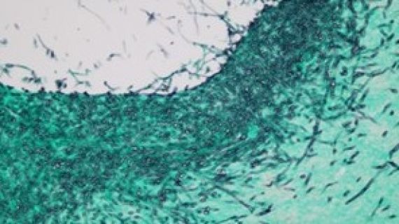
Gomoris Grocott Hexamine (GMS)
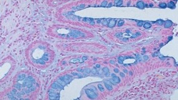
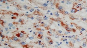
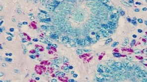
Alcian blue (mast cells)
| Stain Description |
|---|
The Alcian Blue technique is used to demonstrate of mucin and also mast cells. The alcian blue stain is most commonly used on tissue samples obtained from the gastrointestinal (GI) tract This technique can be used in conjunction with other staining methods such as Haematoxylin and Eosin, Van Giesons and Periodic Acid Schiffs. Acidic mucoploysaccharides are stained blue by the Alcian Blue technique, ie mucus, cartilage etc. |
| Results |
| Acidic Muco substances are stained blue |
Congo red
| Stain Description |
|---|
Congo red is used for staining in amyloidosis, and for the cell walls of plants and fungi, and for the outer membrane of Gram-negative bacteria. Apple-green birefringence of Congo red stained preparations under polarized light is indicative of the presence of amyloid fibrils. |
| Results |
| Amlyoid deep red under light microscope. Bright apple green using polarized light. |
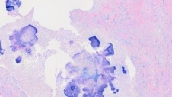
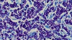
Giemsa Stain
| Stain Description |
|---|
Giemsa method can be used to stain impression smears, blood films and tissue sections. It is commonly used to demonstrate micro-organisms. |
| Results |
| Micro-organisms purplish blue, Cell nuclei blue, Connective tissues pink, Red blood cells salmon pink and Starch and cellulose sky blue. |
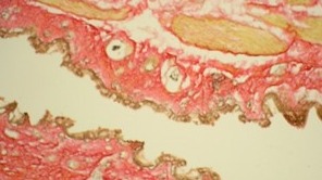
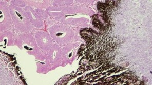
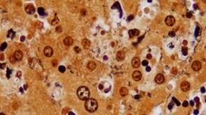
Gomoris Grocott Hexamine (GMS)
| Stain Description |
|---|
The Gomoris Grocott Hexamine Silver technique is used for the demonstration of fungi and basement membrane. The majority of fungi are Periodic Acid Schiffs +ve, but the Gomori”s Hexamine Silver technique is widely used. Some fungi can also be stained by the Gram methods, with hyphae staining Gram positive and spores staining Gram negative. |
| Results |
PAS positive structures, Basement membranes, fungi, Pneumocytis carinii, chitin, glycogen and starch. Black/brown. Mucin Grey to rose. Background Light green |
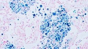
Gram stain


| Stain Description |
|---|
| The Gram stain technique is used to identify and distinguish between Gram- positive and Gram- negative micro-organisms. |
| Results |
Gram positive organisms are deep purple/blue, Gram negative organisms are magenta/pink, and the background is green. |
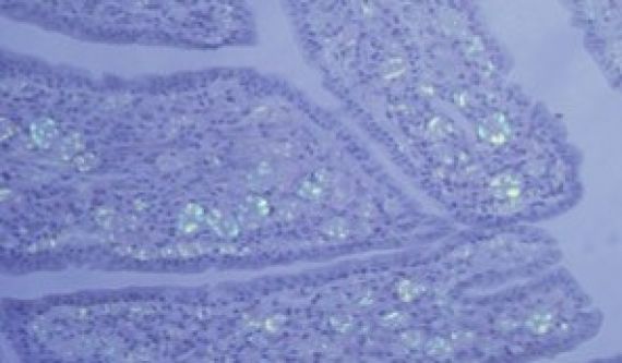
Luxol Fast Blue (LFB)


| Stain Description |
|---|
Luxol fast blue stain (LFB) is used to visualize myelin in nerve tissue. Staining with LFB makes it possible to visualize the myelin and the fatty substance of the brain called white matter which includes the axons of the nerve cells. This stain is often combined with PAS and Haematoxylin. The above two pictures show LFB without counterstain (Blue and white) and LFB with PAS counterstain (Blue and pink). |
| Results |
Myelin and erythrocytes are blue, and the background is pink. |
Massons Trichrome
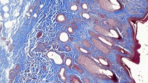
| Stain Description |
|---|
As the name implies three stains (tri) are used selectively staining muscle collagen fibres, fibrin and erythrocytes types of tissue. Standard applications: Masson's trichrome staining is widely used to study muscular pathologies (muscular dystrophy), cardiac pathologies (infarct), hepatic pathologies (cirrhosis) or kidney pathologies (glomerular fibrosis). |
| Results |
Nuclei are purple/black; Muscle fibres, erythrocytes cytoplasm and fibrin are red; and Connective tissue and collagen and green. |
Modified ZN (MZN)
| Stain Description |
|---|
The Modified ZN (MZN) technique is used by this laboratory to positively identify Chlamydia. |
| Results |
Chlamydia comes up as Magenta, Nuclei green and other tissue constituents are green. |
Periodic Acid Schiffs (PAS)
| Stain Description |
|---|
The Periodic Acid Schiff’s Technique is performed by this laboratory for the demonstration of fungi, and carbohydrates particularly glycogen. PAS staining is mainly used for staining structures containing a high proportion of carbohydrate macromolecules (glycogen, glycoprotein, proteoglycans), typically found in e.g. connective tissues, mucus, the glycocalyx, and basal laminae. |
| Results |
Hyphae come up as dark pink. |
Perles Prussian Blue
| Stain Description |
|---|
The Perles Prussian Blue technique is used by this laboratory for the demonstration of Haemosiderin (ferric iron). Deposits of ferric iron in haematoxylin and eosin-stained sections appear as golden green, coarsely to finely granular, intracellular material known as haemosiderin. |
| Results |
Haemosiderin (ferric iron) is blue, and the background/nuclei is red. |
Rhodanine Copper
| Stain Description |
|---|
The Rhodanine technique is used by this laboratory for the demonstration of copper deposits. The reaction utilizes the property of copper to bind with high affinity to protein copper deposits in tissue sections. |
| Results |
Copper comes up as bright red. |
Toluidine Blue
| Stain Description |
|---|
Toluidine blue is used for staining mast cells and Nissli substance. Use of toluidine blue in tissue sections is done with the aim to highlight components, such as mast cells granules, mucins, and cartilage. |
| Results |
Orthochromatic colour, Nissli substance, nuclei and cytoplasm are blue, Metachromic colour is red, and Granules of mast cells are purple. |
Van Giesons
| Stain Description |
|---|
It is the simplest method of differential staining of collagen and other connective tissue. Used often to demonstrate muscle. |
| Results |
Nuclei come up as black/brown; Collagen and connective tissue come up as red; and Cytoplasm, smooth and striated muscle, keratin, erythrocytes come up as yellow. |
Von Kossa
| Stain Description |
|---|
The Von Kossa staining technique is used to stain Calcium salts that are normally present in bone and teeth, and in some pathological conditions, deposits of calcium that are formed in tissues normally devoid of them. |
| Results |
Nuclear chromatin, cytoplasm, collagen comes up as blue/purple; Keratin and erythrocytes come up as pink and Calcium deposits come up as black. |
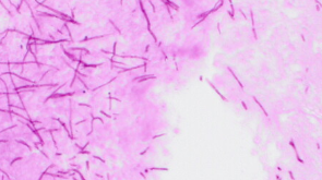
Warthyn Starry
| Stain Description |
|---|
This is a silver technique for the demonstration of spirochaetes in paraffin sections. Warthin-Starry (WS) staining is an ancillary stain used in the detection of Helicobacter sp., spirochaete and other microorganisms in tissue sections. |
| Results |
Spirochaetes and other bacteria come up as black and background tissues come up as yellow/brown. |
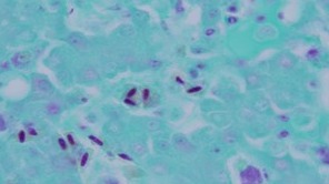
Zeihl Neelson (ZN)
| Stain Description |
|---|
The Ziehl Neelson (ZN) technique is used to positively identify Acid fast mycobacteria. This stain is used as a routine confirmation stain for Johnes disease in sheep and cattle but is also used to identify the presence of Tuberculosis. |
| Results |
Acid-fast bacilli come up as magenta coloured, and Nuclei and other tissue components come up as blue. |
Additional Information

Specimen requirements
| Fixed tissue |
|---|
|
| Paraffin block |
|
| Prepared cassette in formalin |
|
Customer Service
Contact Customer Service for enquiries relating to testing and results, quotations and pricing, couriers, sample submission and invoicing.
NSW DPIRD's laboratories are committed to continual improvement of services. If you would like to provide feedback, please fill in and submit the online feedback form.
CONTACT DETAILS | |
|---|---|
| Phone | 1800 675 623 |
| Operating Hours | 8:30am-4:30pm Monday-Friday (excluding public holidays) 8:30am-12:00pm Saturday (for deliveries only) |
| Postal Address | Private Bag 4008, Narellan NSW 2567 |
Quotations
For Animal export, Plant health or Veterinary quotations, please complete the online quotation request
For Edible oil & oilseed or Feed quality quotations and pricing enquiries, please contact DPIRD AgEnviro Labs at Wagga Wagga on (02) 6938 1957
For Plant nutrition, Soil health or Water quality quotations and pricing enquiries, please contact DPIRD AgEnviro Labs at Wollongbar on (02) 6626 1103
Submission Forms
Veterinary Specimen Advice Form
Please Note: These documents are not fully web accessible, please contact Customer Service (laboratory.services@dpird.nsw.gov.au) for more information.
Courier services and specimen delivery
For NSW submissions, please contact the relevant Customer Service team for courier account details and to order consignment notes.
For submitters outside of NSW, Couriers can be contacted directly to make bookings and arrange collection of packages at the submitters own cost.
Commercial couriers may use either road or air transport and specimens should therefore be packed in accordance with International Air Transport Association (IATA) requirements. Most submission sent by veterinarians to the APHL for testing are defined as “Biological Substance Category B” and must be packed according to the IATA packing instructions 650 (Biological Substance Category B). As requirements for transport can change, customers should consult with their transport agent to obtain current requirements.
Samples may also be hand delivered to the site during normal business hours (8.30am - 4.30pm).
Sample Type | Laboratory | Street Address |
|---|---|---|
Animal (including aquatic animals) | NSW Animal and Plant Health Laboratories (APHL) | EMAI, Woodbridge Road, Menangle NSW 2568 |
