

NSW Animal and Plant Health Laboratories

Our role
The diagnostic laboratory facilities at the NSW Animal and Plant Health Laboratories (formerly known as the State Veterinary Diagnostic Laboratory or SVDL) offer quality assured, comprehensive diagnostic testing and result interpretation for all veterinary species.
NSW Animal and Plant Health Laboratories (APHL) are closely aligned with the NSW Chief Veterinary Officer and the Animal Biosecurity, Biosecurity Policy and Legislation, Animal Welfare, Invasive Plants and Animals and Animal Compliance teams within Biosecurity NSW.
National linkages are maintained through involvement with Animal Health Committee working groups, Australian Animal Pathology Standards Program, Australian Veterinary Association and Animal Health Australia.
NSW APHL is recognised as a pathology training facility by the American College of Veterinary Pathologists and provides pathology training for residents seeking membership by examination through the Australian and New Zealand College of Veterinary Scientists (pathology chapter) or as a Diplomate of the American College of Veterinary Pathology.
Customer service
Contact Customer Service for enquiries relating to testing and results, quotations and pricing, couriers, sample requirements and invoicing.
NSW DPI's laboratories are committed to continual improvement of services. If you would like to provide feedback. please fill in and submit the online feedback form.
Emergency response
NSW APHL provides a critical service in the diagnosis and management of an Emergency Animal Disease (EAD) where the resources of the laboratory are required to be 'emergency-ready' and able to cope with a significant increase in testing.
Suspect EAD can be reported via the Emergency Animal Disease Hotline on 1800 675 888.
To combat emerging disease threats, the NSW APHL continually seeks to improve testing capabilities by upgrading or developing new tests. These improvements are developed and validated in accordance with our NATA quality assurance (QA) requirements.
NSW Animal & Plant Health Laboratories price lists:
The bacteriology laboratory offers a range of routine, anaerobic and selective diagnostic testing, export testing, market assurance and accreditation.
Clinical bacteriology tests include:
- Aerobic and anaerobic culture of bacterial and fungal pathogens from animals
- Antimicrobial susceptibility testing
- Culture of Mycobacterium avium ssp. paratuberculosis (Johnes disease) - herd environment testing (dairy cattle) and individual or pooled animal testing
- PCR tests for slow growing, and difficult-to-culture bacteria
- Detection of C. perfringens enterotoxin
- Strangles exclusion testing via culture and PCR
- Brucella ovis, Actinobacillus seminis and Histophillus somni culture on ram semen
Mastitis bacteriology tests include:
- Culture for clinically significant organisms
- Antimicrobial susceptibility testing for clinical mastitis cases
- Routine herd screenings and bulk-tank milk testing
Bee Diagnostics:
- American foulbrood (AFB) and European (EFB) identification in diseased larvae
- AFB culture on honey
Collection of samples for bacterial testing should be as aseptically as possible to reduce culture overgrowth with contaminants. Find out more about the range of available tests and for information on how to collect, package and submit samples for bacteriology testing refer to the veterinary test list.
The Biotechnology laboratory performs molecular based testing for genetic disease, genetic traits as well as species identification for a range of reasons including;
- Disease diagnosis
- Disease management
- Emergency response
- Registration in breed societies
- Entry to AI centres
- Export and import of animals or semen
- Sale of animals
- Herd/flock management
DNA testing (genotyping) provides a tool for producers to identify animals with favourable and unfavourable alleles to facilitate effective management.
Sample types include hair, unclotted blood (EDTA or Lithium Heparin tubes), semen, tissue, ear notches, faeces or DNA from other testing facilities. All samples are tested via PCR;
- Real-time PCR on all high-throughput tests
- Conventional gel-based PCR on low volume tests
To view the range of available tests and for information on how to collect, package and submit samples refer to the veterinary test list.
The histology laboratory prepares slides for microscopic examination which supports the team of dedicated veterinary pathologists to observe and interpret morphological changes to animal tissue. Tissue specimens are generally collected from post mortems (field or in-house) or biopsies from live animals.
The process of producing a slide for microscopic examination generally involves sample processing, embedding, sectioning, staining and cover slipping.
Prompt fixation of samples in 10% neutral buffered formalin to preserve the tissue is crucial, particularly with large whole organs such as brain tissue. Inadequate fixation will result in autolysis or post mortem degeneration which reduces the ability to produce quality slides for histological examination.
The histopathology laboratory also offers quality digital scanning of physical glass slides with the ability to securely store images on the internet enabling global collaboration and sharing. Digital scanning of glass slides assists in being able to quickly identify and respond to biosecurity threats with the ability to collaborate nationally and globally with specialist veterinary pathologists.
Find out more about how to collect, package and submit samples for histopathology testing.
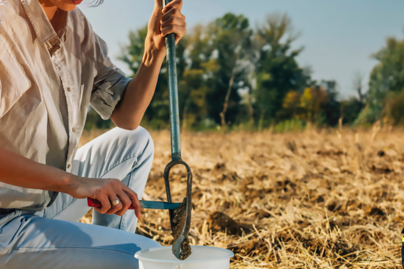
The parasitology laboratory provides diagnostic testing on production, performance and companion animals. Parasitological techniques used by this laboratory include parasite identification, Baermann for lungworm or Strongyloides, quantitative faecal examination and total worm counts of the gastrointestinal tract (GI) or GI washings.
WormTest Kits provided by NSW DPI can be used as a simple and quick way to monitor your worm control program by checking drench effectiveness.
The parasitology laboratory also offers worm typing in addition to the WormTest kits. Faecal samples are pooled for culturing of worm eggs. The larvae which hatch are examined and identified. Due to the time require to culture and differentiate, worm typing results take approximately ten days.
WormTest results can be used to help determine when to treat your animals. Drenching when it is not necessary may promote development of resistance to the worming compound among those worms that are present. Worm testing is also useful if you suspect that current worming treatments are not working effectively.
If you are unsure what type of WormTest to choose we encourage you to consult with a veterinarian or other suitably qualified adviser.
WormTest Kits are available from rural suppliers or from NSW DPI upon request. Each kit contains an information sheet, collection instructions, prepaid postage to the laboratory, a mailing container, a glove and ten sample containers. To request a WormTest Kit contact Customer Service.
Find out more about how to collect, package and submit samples for parasitology testing.
The SVDL has a team of dedicated and highly qualified pathologists trained in the diagnostic evaluation and interpretation of terrestrial and aquatic diseases. Case management is undertaken as required with a dedicated pathologist assigned to investigate ongoing or emerging diseases. A collaborative team approach ensures that advice on testing requirements, differential diagnoses and result interpretation is readily available and consistently delivered.
The serology laboratory is a high volume laboratory providing services for diagnostic testing, export testing, AI entry, market assurance and accreditation.
Tests performed by the Serology laboratory include:
- Complement Fixation Test (CFT)
- Enzyme-linked immunoabsorbant assay (ELISA)
- Microscopic Agglutination Test (MAT)
- Serum Agglutination Test (SAT)
- Rapid Plate Tests
- Clinical Pathology Tests
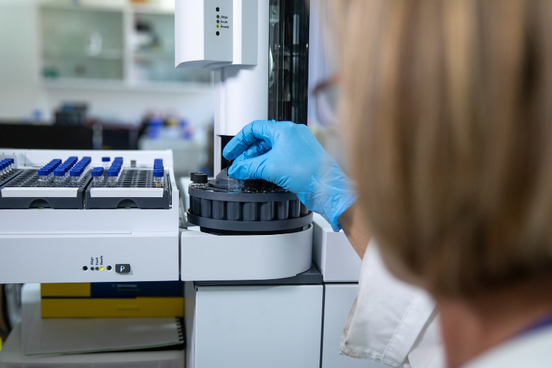
The Serology laboratory uses robotics to process samples for testing, ensuring a faster turnaround time.
Fresh blood samples are suitable for serology tests. Ideally samples should be submitted to the laboratory under refrigeration. Samples that are haemolysed due to exposure to hot conditions are not suitable for testing.
Blood tubes submitted for serology testing should not be sealed with sticky tape or have protruding labels. It is recommended that a key list accompany each batch of serology samples with the number written on each tube with a thick waterproof pen.
To view the range of available tests and for information on how to collect, package and submit samples refer to the veterinary test list.
The veterinary virology laboratory provides diagnostic and research services for viral diseases of commercial livestock, wildlife and aquatic animal species.Testing is conducted Monday to Friday. The laboratory has a team of veterinary virologists to support these services.
Viruses are detected through the testing of tissues, serum, faeces, and other samples using a variety of specialised techniques, including electron microscopy.
The veterinary virology laboratory service includes antigen/nucleic acid/antibody detection tests for all of the common mammalian and avian viruses, and selected aquatic viruses
Research programs have been developed cooperatively with industry to investigate significant diseases in livestock and make recommendations on effective control programs. The major emphasis is on identification of new and emerging viral diseases, development of improved diagnostic tests and vaccine production, and certification testing to facilitate overseas trade. Major programs currently involve Pestivirus in all animal species (the Virology Laboratory at EMAI is a World Reference Laboratory for Pestiviruses) and arboviruses in cattle and sheep.
To view the range of available tests and for information on how to collect, package and submit samples refer to either the Virology Test Page, or the A-Z Veterinary Test List.
